Faster Diagnoses, Lower Costs, Better Care
Medical Disclaimer: This article is for informational and educational purposes only and does not constitute medical advice, diagnosis, or treatment. The technologies, applications, and outcomes described reflect general industry trends and published research; individual patient experiences will vary based on clinical circumstances, the specific tools deployed at a given facility, and the judgment of treating physicians. AI in radiology is intended to assist licensed radiologists, not replace them. Readers should not make medical decisions based on this article and should consult qualified healthcare providers for any medical questions or concerns. If you are experiencing a medical emergency such as stroke symptoms — sudden weakness, facial drooping, confusion, or difficulty speaking — call 911 immediately.
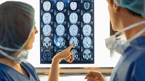
If you’ve had a CT scan, MRI, or mammogram in Florida in the last two years, there’s a reasonable chance an artificial intelligence system looked at the images before — or alongside — the radiologist who signed your report. You probably weren’t told. The technology runs in the background, flagging abnormalities, prioritizing urgent cases, and helping radiologists work through ever-larger imaging volumes. Done well, it makes diagnoses faster, more accurate, and meaningfully less expensive.
Florida sits in the middle of this shift. The state has a large, aging population, a sprawling network of imaging centers and hospital systems, and persistent radiologist staffing pressures — exactly the conditions where AI offers the most concrete value. Here’s what it actually does, where it’s deployed, and what it means for Florida patients.
The Scale of What’s Happening
As of late 2025, the FDA had cleared more than 1,300 AI-enabled medical devices. Roughly 76–80% of them — over 1,000 tools — are radiology applications. That makes radiology, by a wide margin, the most AI-saturated specialty in medicine. And the pace is accelerating: the FDA added 56 radiology-specific AI tools in a single update in late 2025.
These aren’t experimental. They’re software products integrated into the imaging systems and reporting workflows already running at hospitals and outpatient imaging centers across Florida.
What AI Actually Does in a Radiology Department
Most patients picture AI as something that “reads” their scan and produces a diagnosis. The reality is more layered. AI in radiology generally falls into six functional buckets:
1. Worklist prioritization. When a scan suggests a stroke, pulmonary embolism, brain bleed, or collapsed lung, AI flags it and pushes it to the top of the radiologist’s queue — sometimes within seconds of image acquisition. This is the single highest-value application in emergency settings.
2. Detection and characterization. Algorithms highlight suspicious areas: lung nodules on chest CTs, microcalcifications on mammograms, fractures on X-rays, vessel occlusions on CT angiograms, lesions on abdominal scans. The radiologist still makes the call; the AI ensures nothing obvious is overlooked.
3. Quantification. AI measures things faster and more consistently than humans can — tumor size over time, brain volume changes in suspected dementia, breast density, coronary calcium scores, organ volumetrics. Consistency matters when tracking a patient across years of scans.
4. Image enhancement. Newer algorithms reconstruct higher-quality images from lower-dose CT scans, shorter MRI sequences, or smaller contrast doses. For Florida’s large senior population, lower radiation exposure and reduced contrast — which can stress kidneys — is a meaningful safety improvement.
5. Workflow automation. Drafting structured report text, scheduling, predicting no-shows, optimizing scanner protocols. None of this touches a diagnosis directly, but it frees radiologist time for the cases that need it most.
6. Triage and care coordination. Platforms like Viz.ai, used by more than 1,700 hospitals nationwide, automatically alert stroke or PE response teams the moment a scan shows a treatable problem — often before the radiologist has even opened the study.
How AI Improves Diagnostic Accuracy
The published evidence varies by application, but several findings are consistent:
- AI-assisted detection of large vessel occlusion in stroke shows up to 95% sensitivity in published studies.
- Lung nodule detection on chest CT regularly exceeds 94% segmentation accuracy.
- Reading times drop 30–50% across many high-volume study types.
- Scan times themselves drop 30–75% with AI-accelerated MRI reconstruction.
For stroke specifically, AI has changed the standard of care. American Heart Association/American Stroke Association guidelines now recognize AI-augmented stroke imaging as Level 1 evidence, and the Centers for Medicare & Medicaid Services issued the first reimbursement for AI-augmented stroke care back in 2020. The clinical reasoning is straightforward: in stroke, every minute of delay costs roughly 1.9 million neurons. AI that shaves 15–30 minutes off the path from scan to treatment translates directly to preserved brain function.
It’s worth being honest about the evidence gaps. A 2024 systematic review of 950 FDA-authorized AI/ML devices found that only about 5% of radiology AI devices underwent prospective testing before clearance, and only 29% incorporated clinical testing in their submissions. Most cleared the FDA’s 510(k) pathway by demonstrating equivalence to existing devices rather than head-to-head superiority trials. That doesn’t mean these tools don’t work — many demonstrably do — but it does mean radiologists, not AI, remain the accountable decision-makers, and clinical oversight matters.
How AI Reduces Costs
The cost story has multiple threads, some well-documented and some still emerging.
Stroke care. A peer-reviewed cost-effectiveness analysis modeling AI-aided detection of large vessel occlusion found that, with realistic assumptions about missed-diagnosis rates and AI performance, the technology produced both cost savings and improved quality-adjusted life years. For one annual cohort of suspected stroke patients in the UK, modeled savings reached approximately $11 million. Florida’s stroke volumes — driven by the state’s senior population — are far higher, and the per-patient logic translates.
Preventing the high cost of missed diagnoses. The most expensive scan is the one read incorrectly. A missed lung cancer caught at stage IV instead of stage I, a delayed stroke that costs a patient months of rehabilitation, an undetected pulmonary embolism that ends in cardiac arrest — these are the events AI is best positioned to prevent, and they dwarf the per-scan cost of the software.
Throughput. Florida imaging centers are experiencing the same radiologist staffing crunch as the rest of the country. AI that lets a radiologist read 20–30% more studies per shift effectively expands capacity without expanding headcount — keeping wait times shorter and per-study overhead lower.
Lower-dose imaging. AI-reconstructed images allow lower radiation doses on CT and shorter MRI scan times. Less time on the scanner means more patients per machine per day, lower contrast costs, and reduced downstream costs from radiation-related morbidity over a patient’s lifetime.
Reduced unnecessary follow-up. Better quantification and consistent measurements reduce the “is it growing or am I imagining it?” follow-up scans that drive imaging spending.
What hasn’t been fully resolved is reimbursement. Outside a handful of approved use cases (stroke being the most prominent), insurance reimbursement for AI-aided reads is still rudimentary. Most Florida hospitals absorb AI software costs as an operational expense, betting on workflow efficiency and risk reduction rather than direct billing.
Who’s Using It in Florida
AI-enabled imaging is now standard at most major Florida health systems. While specific deployments vary and change frequently, the institutions investing publicly in radiology AI include:
- Tampa General Hospital has hosted dedicated AI innovation events featuring partners like Agfa HealthCare’s RUBEE Augmented Intelligence platform, and operates one of the larger interventional radiology programs in the state.
- AdventHealth Central Florida operates more than 100 radiologists across eight subspecialties, with imaging centers across the I-4 corridor and ongoing AI integration in mammography and cross-sectional imaging.
- HCA Florida Healthcare — the state’s largest hospital system — runs ACR-accredited imaging across dozens of facilities with intelligent dose management and AI-assisted CT protocols.
- Orlando Health integrates AI tools across its system imaging departments.
- Baptist Health South Florida, Cleveland Clinic Florida, University of Miami / Bascom Palmer, UF Health Shands, and Mayo Clinic Jacksonville all run substantial radiology AI programs.
- Specialty Focused Radiology (Tampa-based) and Radiology Associates of Florida are among the larger radiology groups deploying AI-assisted reads across hospital and outpatient settings statewide.
Independent imaging centers, large radiology groups, and teleradiology providers serving rural Florida hospitals are increasingly adopting AI as well — particularly for stroke triage, where 24/7 expert coverage is hardest to staff.
What This Means for Florida Patients
For most patients, AI in radiology will be invisible. You’ll get a scan; the report will arrive faster; if something urgent is found, the response will start sooner. A few practical implications worth knowing:
- AI doesn’t replace your radiologist. Every cleared tool in clinical use today is decision-support software. A board-certified radiologist still interprets and signs your report.
- You can ask whether AI was used. It’s a reasonable question. Larger systems are increasingly transparent about which tools they use.
- AI works best with prior imaging. If you’ve had scans elsewhere, getting them transferred to your current imaging center improves both AI and human accuracy. Most quantitative AI tools rely on comparison with prior studies.
- Stroke symptoms are still a 911 call. No AI tool helps if you don’t get to the scanner in the first place. Sudden weakness on one side, facial drooping, slurred speech, severe headache, or vision loss require immediate emergency care.
What’s Coming
Several developments are likely to shape Florida radiology over the next two to three years:
- Generative AI in reporting. Large language models drafting structured radiology reports from imaging findings are moving from prototype to deployed product. Expect faster turnaround and more consistent reports, with radiologists editing rather than typing from scratch.
- Population-scale screening. AI lowers the marginal cost of reading scans enough that broader screening programs — for lung cancer, coronary calcium, osteoporosis — become economically viable. Florida’s senior population is a natural target.
- Regulatory evolution. The FDA is piloting “predetermined change control plans” that let manufacturers update AI models without full re-clearance, acknowledging that AI evolves over time. The EU AI Act, effective January 2026, classifies medical AI as high-risk and imposes stricter validation requirements; U.S. policy is likely to move in a similar direction.
- Reimbursement codes. Expect more CPT codes for AI-augmented imaging in the next two to three years, which will accelerate adoption at smaller facilities that currently can’t justify the software cost.
The Bottom Line
AI in radiology isn’t a future promise in Florida — it’s deployed, in production, reading studies right now at most of the imaging centers and hospitals you’d be referred to. The evidence is strongest for time-critical applications like stroke and pulmonary embolism, solid for cancer screening and follow-up, and still maturing for many other use cases. Costs are coming down, accuracy is going up, and radiologists — far from being replaced — are using AI to handle the steadily growing volume of imaging that Florida’s healthcare system needs interpreted every day.
For patients, the practical takeaway is simple: the technology is making the system around your care faster and more accurate, but the relationship that matters is still the one between you, your physician, and the radiologist reading your scan.
Disclaimer (repeated): This article does not provide medical advice, diagnosis, or treatment recommendations. References to specific hospitals, vendors, products, and platforms are for informational purposes only and do not constitute endorsement. AI capabilities, FDA clearances, insurance coverage, and institutional deployments described here may have changed since publication. Patients should consult their physicians and radiologists about how imaging is interpreted at the facility serving them. If you are experiencing what may be a medical emergency, call 911 or go to the nearest emergency department immediately.
Stroke Warning Signs (BE FAST): Balance loss, Eye/vision changes, Face drooping, Arm weakness, Speech difficulty, Time to call 911. Stroke care is one of the most time-sensitive applications of imaging AI — but only if patients reach the hospital in time.